Smooth Muscle Diagram Labeled / Skeletal Muscle Tissue Diagram Labeled Instituto / Skeletal muscles attach to and move bones by contracting and relaxing in response to voluntary messages from the nervous system.
Smooth Muscle Diagram Labeled / Skeletal Muscle Tissue Diagram Labeled Instituto / Skeletal muscles attach to and move bones by contracting and relaxing in response to voluntary messages from the nervous system.. Skeletal muscles attach to and move bones by contracting and relaxing in response to voluntary messages from the nervous system. In skeletal muscle, a single type of somatic nervous system traverses to muscle, where it stimulates organelle in the muscle cells in order to release calcium. Diagram of smooth muscle cardiac muscle and straited muscle tissue. Structure and composition of muscle meat science. On the left is the view with light microscopy.
Smooth muscle is composed of sheets or strands of smooth muscle cells. Smooth muscle is made up of cells that contain a single central nucleus. 1 response to simple smooth muscle diagram labeled unknown june 5, 2021 at 12:39 am. (muscle cells are often referred to as muscle fibers because of their narrowness and length.). Which of the labeled layers in the diagram of the arterial wall is composed of a simple squamous epithelium, a basement membrane and a layer of elastic tissue?
Draw A Labelled Diagram Of Cardiac Muscle Found In Human Body Write Two Differences Between Striated And Smooth Muscles Science Tissues 9476045 Meritnation Com from s3mn.mnimgs.com Diagram of smooth muscle cardiac muscle and straited muscle tissue. Smooth muscles are unique in their largely involuntary response, and in their structure. It is the pen diagram of skeletal, smooth and cardiac muscle for class 10, 11 and 12. Diagram of smooth muscle contraction, smooth cardiac and skeletal muscle diagram, smooth muscle cell diagram, smooth muscle cell picture. This set contains the following 10 diagrams: On the left is the view with light microscopy. Human anatomy muscle organ diagrams by julie ridge are part of my human anatomy series. Smooth muscle tissue, unlike striated muscle, contracts slowly and automatically.
In skeletal muscle, a single type of somatic nervous system traverses to muscle, where it stimulates organelle in the muscle cells in order to release calcium.
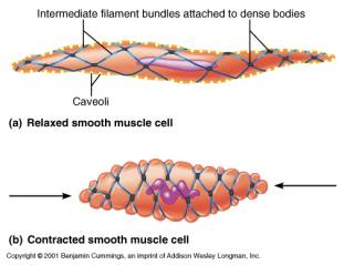
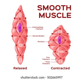
This diagram shows a few of the cells that can be seen in the stained section below. In this video i have shown the simplest way of drawing muscle drawing. Blood vessels contain only smooth muscle cells. Name three types of fiber arrangements seen in skeletal muscle. This set contains the following 10 diagrams: The muscles of the human body can be categorized into a number of groups which include muscles relating to the head and neck, muscles of the torso or trunk, muscles of the upper limbs, and muscles of the lower limbs. Diagram of smooth muscle contraction, smooth cardiac and skeletal muscle diagram, smooth muscle cell diagram, smooth muscle cell picture. You will also get the identification points of skeletal muscle histology slide with a little description here in this guide. They range from about 30 to 200 μm (thousands of times shorter than skeletal muscle fibers), and they produce their own connective tissue, endomysium.although they do not have striations and sarcomeres, smooth muscle fibers do have actin and myosin. These muscle cells reside within the tunica media along with elastic fibers and connective tissue. Skeletal muscles attach to and move bones by contracting and relaxing in response to voluntary messages from the nervous system. Smooth muscle is found in the walls of hollow organs like your intestines and stomach. Muscle anatomy coloring sheets 12 photos of the muscle anatomy coloring sheets free muscle anatomy coloring sheets, muscle anatomy coloring pages, muscle anatomy coloring pages free, muscle anatomy coloring sheets, human muscles, free muscle anatomy coloring sheets, muscle anatomy coloring pages, muscle anatomy.
Diagram of smooth muscle contraction, smooth cardiac and skeletal muscle diagram, smooth muscle cell diagram, smooth muscle cell picture. Individual muscle fibers, (b) surrounds groups of skeletal muscle fibers (fascicles), and (c) covers the muscle as a whole. Which of the labeled layers in the diagram of the arterial wall consists mainly of elastic fibers and smooth muscle fibers? They range from about 30 to 200 μm (thousands of times shorter than skeletal muscle fibers), and they produce their own connective tissue, endomysium.although they do not have striations and sarcomeres, smooth muscle fibers do have actin and myosin. Related posts of smooth muscle diagram labeled muscle anatomy coloring sheets.
Histology Of Muscle from faculty.etsu.edu Human anatomy muscle organ diagrams by julie ridge are part of my human anatomy series. Smooth muscle is made up of cells that contain a single central nucleus. Smooth muscle anatomy smooth muscle tissue is also known as visceral muscle tissue. Its wavelike movements propel things through the bodily system, such as food through. Smooth muscle, muscle that shows no cross stripes under microscopic magnification. Muscle fiber diagram showing the muscle, fascicle, muscle fibers, myofibril, and Diagram of smooth muscle contraction, smooth cardiac and skeletal muscle diagram, smooth muscle cell diagram, smooth muscle cell picture. A muscle cell is a long cell as compared to other kinds of cells, and many muscle cells connect with each other to create the long fibers present in muscle tissue.
Smooth muscle, muscle that shows no cross stripes under microscopic magnification.
The single muscle cell consists of many nuclei that are pressed against the cell membrane. Which of the labeled layers in the diagram of the arterial wall consists mainly of elastic fibers and smooth muscle fibers? Its wavelike movements propel things through the bodily system, such as food through. The innermost layer of the stomach wall is the gastric mucosa.it is formed by a layer of surface epithelium and an underlying lamina propria and muscularis mucosae. This smooth muscle can be found surrounding the walls of the blood vessels, the bronchioles in the lungs, and the sphincter muscles used in the gi tract.the gi tract, which is tubular by design, also houses longitudinal muscles in addition to the smooth. Smooth muscles are involved in many. Diagram of contraction of skeletal muscle. Smooth muscles are unique in their largely involuntary response, and in their structure. The surface epithelium is a simple columnar epithelium.it lines the inside of the stomach as surface mucous cells and forms numerous tiny invaginations, or gastric pits, which appear as millions of holes all throughout the stomach. 1 response to simple smooth muscle diagram labeled unknown june 5, 2021 at 12:39 am. Skeletal muscle tissue is composed of long cells called muscle fibers that have a striated appearance. The cells stick together and are connected by specialised cell junctions, called gap junctions. Smooth muscle is made up of cells that contain a single central nucleus.
In this short guide, you will get a basic concept of skeletal muscle histology from the real slide and labeled diagram. Muscle anatomy coloring sheets 12 photos of the muscle anatomy coloring sheets free muscle anatomy coloring sheets, muscle anatomy coloring pages, muscle anatomy coloring pages free, muscle anatomy coloring sheets, human muscles, free muscle anatomy coloring sheets, muscle anatomy coloring pages, muscle anatomy. Smooth muscles are involved in many. Smooth muscle fibers are often found forming sheets of tissue and function in a coordinated fashion due to the presence of gap junctions between the cells. Smooth muscle, muscle that shows no cross stripes under microscopic magnification.
Smooth Muscle Hd Stock Images Shutterstock from image.shutterstock.com This smooth muscle can be found surrounding the walls of the blood vessels, the bronchioles in the lungs, and the sphincter muscles used in the gi tract.the gi tract, which is tubular by design, also houses longitudinal muscles in addition to the smooth. On the left is the view with light microscopy. A muscle cell is a long cell as compared to other kinds of cells, and many muscle cells connect with each other to create the long fibers present in muscle tissue. Muscle anatomy coloring sheets 12 photos of the muscle anatomy coloring sheets free muscle anatomy coloring sheets, muscle anatomy coloring pages, muscle anatomy coloring pages free, muscle anatomy coloring sheets, human muscles, free muscle anatomy coloring sheets, muscle anatomy coloring pages, muscle anatomy. Smooth muscle (textus muscularis levis) smooth muscle is a type of tissue found in the walls of hollow organs, such as the intestines, uterus and stomach. Smooth muscle diagram labeled tag smooth muscle tissue labeled diagram human anatomy diagram, picture of smooth muscle diagram labeled tag smooth muscle tissue labeled diagram human anatomy diagram Muscle fiber diagram showing the muscle, fascicle, muscle fibers, myofibril, and Blood vessels contain only smooth muscle cells.
Individual muscle fibers, (b) surrounds groups of skeletal muscle fibers (fascicles), and (c) covers the muscle as a whole.
Which of the labeled layers in the diagram of the arterial wall is composed of a simple squamous epithelium, a basement membrane and a layer of elastic tissue? Name three types of fiber arrangements seen in skeletal muscle. In this video i have shown the simplest way of drawing muscle drawing. Smooth muscle fibers are often found forming sheets of tissue and function in a coordinated fashion due to the presence of gap junctions between the cells. Although vessels only contain smooth muscles, the contraction of skeletal muscle plays an important role in the movement of blood from the periphery towards the heart in the venous system. You will also get the identification points of skeletal muscle histology slide with a little description here in this guide. Skeletal muscles, smooth muscles, cardiac muscles. This set contains the following 10 diagrams: Smooth muscle is a type of muscle tissue which is used by various systems to apply pressure to vessels and organs. Smooth muscles are unique in their largely involuntary response, and in their structure. Smooth muscle contracts under certain stimuli as atp is freed. Smooth muscle is composed of sheets or strands of smooth muscle cells. Diagram of smooth muscle contraction, smooth cardiac and skeletal muscle diagram, smooth muscle cell diagram, smooth muscle cell picture.
This smooth muscle can be found surrounding the walls of the blood vessels, the bronchioles in the lungs, and the sphincter muscles used in the gi tractthe gi tract, which is tubular by design, also houses longitudinal muscles in addition to the smooth smooth muscle diagram. Diagram of smooth muscle contraction, smooth cardiac and skeletal muscle diagram, smooth muscle cell diagram, smooth muscle cell picture.


0 Komentar